Thursday, November 30, 2006

Frontopolar Cortex Moves to the Back of the Brain

from Christoff & Gabrieli 2000

In case you missed this announcement, as I did, New Scientist has now declared that frontopolar cortex is in the back of the brain:
In addition, unlike in humans, the researchers also found spindle cells in the frontopolar cortex at the back of the brain...
It seems that writers there also expect spindle neurons to be the next new thing.
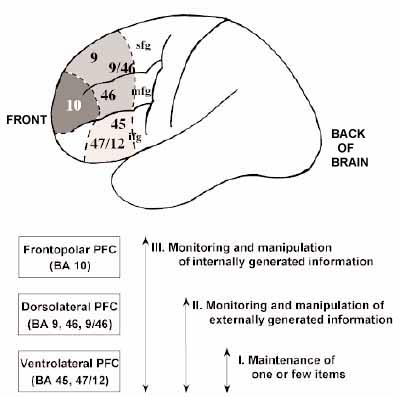
Whales boast the brain cells that 'make us human'
15:00 27 November 2006
Andy Coghlan

. . .

They were touted as the brain cells that set humans and the other great apes apart from all other mammals. Now it has been discovered that some whales also have spindle neurons – specialised brain cells that are involved in processing emotions and helping us interact socially.

Spindle cells, named after their long, spindle-shaped bodies, are the cells that are credited with allowing us to feel love and to suffer emotionally...

The cells occur in parts of the human brain that are thought to be responsible for our social organisation, empathy, speech, intuition about the feelings of others, and rapid "gut" reactions.

. . .

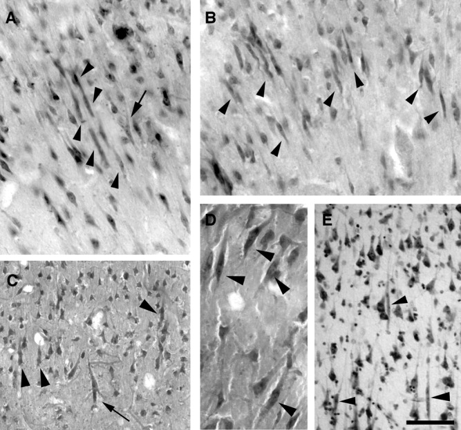
As with humans, the spindle cells were found in whales in the anterior cingulate cortex and frontoinsular cortex – two brain regions vital for "visceral" reactions. Such reactions require fast but emotionally-sensitive judgments, such as deciding whether another animal is suffering pain, and the general feel of whether an experience is pleasant or unpleasant.

In addition, unlike in humans, the researchers also found spindle cells
in the frontopolar cortex at the back of the brain, and they were sparsely dispersed elsewhere...
Hmm, but some researchers have argued that frontopolar cortex (aka Brodmann area 10) is one brain region that allows us to engage in the type of complex thought that distinguishes humans from other animals, described variously as subgoal processing (Braver & Bongiolatti, 2002), complex reasoning (Bunge et al., 2005), and manipulation of internally generated information:
Christoff K, Gabrieli JDE. (2000). The frontopolar cortex and human cognition: evidence for a rostrocaudal hierarchical organization within the human prefrontal cortex. Psychobiology 28: 168-186.

Numerous brain lesion and functional neuroimaging studies have suggested that the dorsolateral and frontopolar prefrontal regions are involved in complex cognitive processes subserving thought and memory. However, previously proposed functional subdivisions of prefrontal function have concentrated predominantly on posterior prefrontal cortex, including the dorsolateral, ventral, and medial regions. Far less consideration has been given to characterizing the psychological processes mediated by the frontopolar cortex. Here we review published neuroimaging studies of reasoning and episodic memory, two domains in which the frontopolar cortex has been frequently activated. The results suggest that dorsolateral prefrontal cortex is involved when externally generated information is being evaluated, whereas the frontopolar cortex becomes recruited when internally generated information needs to be evaluated. A hierarchical model of prefrontal function is proposed in which dorsolateral and frontopolar regions are serially recruited as a reasoning or memory task requires evaluation of internally generated information.
But Hof and Van Der Gucht (2006) carefully qualify the significance of their frontopolar finding in whales:
In addition, spindle cells in Megaptera occur in regions where they had not been seen in hominids such as the frontal polar cortex, although there is likely no functional or topographic homology between the frontopolar region in mysticetes and hominids.
And they know that frontopolar cortex is in the front of the brain.

References

Braver TS, Bongiolatti SR. (2002). The role of frontopolar cortex in subgoal processing during working memory. Neuroimage 15: 523-536.

Bunge SA, Wendelken C, Badre D, Wagner AD. (2005). Analogical reasoning and prefrontal cortex: evidence for separable retrieval and integration mechanisms. Cereb Cortex 15: 239-249.

Hof PR, Van Der Gucht E. (2007). Structure of the cerebral cortex of the humpback whale, Megaptera novaeangliae (Cetacea, Mysticeti, Balaenopteridae). Anatom Rec Part A, 290:1-31.

Subscribe to Post Comments [Atom]

Tuesday, November 28, 2006

Spindle Neurons in Humpback Whales

from Hof & Van Der Gucht (2006)

Spindle Neurons: The Next New Thing?

In July, The Neurocritic wrote about a special class of cells found in the anterior cingulate cortex and frontoinsular cortex of humans and great apes. These spindle neurons are a unique type of neuron found in layer Vb in the ACC and FI cortex of humans. In 1999, it was discovered that besides humans, only great apes have spindle neurons (Nimchinsky et al.).

And now we can add humpback whales to the select group of animals who possess these magical spindle neurons.
Hof PR, Estel Van Der Gucht E. (2006). Structure of the cerebral cortex of the humpback whale, Megaptera novaeangliae (Cetacea, Mysticeti, Balaenopteridae). Anatom Rec Part A, Early View (Published Online: 27 Nov 2006).

Cetaceans diverged from terrestrial mammals between 50 and 60 million years ago and acquired, during their adaptation to a fully aquatic milieu, many derived features, including echolocation (in odontocetes), remarkable auditory and communicative abilities, as well as a complex social organization. Whereas brain structure has been documented in detail in some odontocetes, few reports exist on its organization in mysticetes. We studied the cerebral cortex of the humpback whale (Megaptera novaeangliae) in comparison to another balaenopterid, the fin whale, and representative odontocetes. We observed several differences between Megaptera and odontocetes, such as a highly clustered organization of layer II over the occipital and inferotemporal neocortex, whereas such pattern is restricted to the ventral insula in odontocetes. A striking observation in Megaptera was the presence in layer V of the anterior cingulate, anterior insular, and frontopolar cortices of large spindle cells, similar in morphology and distribution to those described in hominids, suggesting a case of parallel evolution. They were also observed in the fin whale and the largest odontocetes, but not in species with smaller brains or body size. The hippocampal formation, unremarkable in odontocetes, is further diminutive in Megaptera, contrasting with terrestrial mammals. As in odontocetes, clear cytoarchitectural patterns exist in the neocortex of Megaptera, making it possible to define many cortical domains. These observations demonstrate that Megaptera differs from Odontoceti in certain aspects of cortical cytoarchitecture and may provide a neuromorphologic basis for functional and behavioral differences between the suborders as well as a reflection of their divergent evolution.
John Allman's Lab at Cal Tech thinks
they participate in fast, intuitive social decision-making. We have found that the Von Economo [spindle] neurons emerge mainly in the first three years after birth. We also have evidence that in autistic subjects the Von Economo neurons are abnormally located, possibly as a result of a migration defect. This abnormality may be at least partially responsible for defective social intuition in autism.
It's interesting to see how the popular press is covering this finding:
Humpback whales have "human" brain cells: study

WASHINGTON (Reuters) - Humpback whales have a type of brain cell seen only in humans, the great apes, and other cetaceans such as dolphins, U.S. researchers reported on Monday.

This might mean such whales are more intelligent than they have been given credit for, and suggests the basis for complex brains either evolved more than once, or has gone unused by most species of animals, the researchers said.

The finding may help explain some of the behaviors seen in whales, such as intricate communication skills, the formation of alliances, cooperation, cultural transmission and tool usage, the researchers report in The Anatomical Record.

Patrick Hof and Estel Van der Gucht of the Department of Neuroscience at Mount Sinai School of Medicine in New York studied the brains of humpback whales and discovered a type of cell called a spindle neuron in the cortex, in areas comparable to where they are seen in humans and great apes.

Although the function of spindle neurons is not well understood, they may be involved in cognition -- learning, remembering and recognizing the world around oneself. Spindle cells may be affected by Alzheimer's disease and other debilitating brain disorders such as autism and schizophrenia.
Oh, here's a great one. Whale-human chimeras:
New study finds some whales have human brain cells

NEW YORK: Brains of some species of whales have spindle neurons, the elongated nerve cells found in humans and some great apes and associated mostly with decision-taking and involved in higher cognitive functions like consciousness and expressing emotions, say U.S. scientists who have studied whales.

What is surprising is that these cells in whales were found located in the same region as in human brain and these were absent in smaller-brained whales and also in dolphins. Two of the whales which possessed spindle neurons are the humpback whale and the fin whale.

. . .

The two wrote in the report, "In spite of the relative scarcity of information on many cetacean species, it is important to note in this context that sperm whales, killer whales, and certainly humpback whales, exhibit complex social patterns that included intricate communication skills, coalition-formation, cooperation, cultural transmission and tool usage."

Subscribe to Post Comments [Atom]

I'm a C-List Blogger!

Darn! I was hoping to be on the D-List, along with Kathy Griffin.


Kathy with Clay Aiken at the 2004 American Music Awards


C-List Blogger

The Middle Authority Group [C-List Bloggers]
(10-99 blogs linking in the last 6 months)
This contrasts somewhat with the second group, which enjoys an average age not much older than the first at 260 days and which posts 50% more frequently than the first. There is a clear correlation between posting volume and Technorati authority ranking.


Are You an A-List Bloglebrity?

hat tip to Retrospectacle

Subscribe to Post Comments [Atom]

Sie Ist Gefundene Steuerung Über Two Languages: Part 2

In the last post, The Neurocritic examined an fMRI experiment by Crinion et al. (2006) that utilized a semantic priming design to examine language switching in fluent bilinguals. It turned out that activity in a subcortical region in the basal ganglia, or more specifically, the head of the caudate nucleus in the left hemisphere, showed a semantic priming effect when pairs of words were in the same language, but not in different languages:

prime-target pairs ... were either semantically related (bathtub-SHOWER) or unrelated (spoon-SHOWER). We then identified language-dependent semantic responses by comparing the effect of semantic priming when prime and target were in the same language (trout-SALMON in English or forelle-LACHS in German) or different languages (e.g., trout-LACHS or forelle-SALMON).

The time between the onset of the prime word and the onset of the target word was short: 250 msec. This short interval skews semantic access to processes that are more automatic and less "controlled"or predictive (i.e., you don't have time to generate SHOWER as a semantic neighbor to bathtub in less than 250 msec). So the experiment was measuring relatively automatic semantic priming processes. We don't know what would happen if a long prime-target interval was used in order to tap controlled or attentional or predictive processes.

The graph to the left indicates that bathtub-SHOWER (S, semantically related) pairs elicit less activity than spoon-SHOWER (U, unrelated) pairs. But when the prime and target are in different languages, there is no semantic priming effect. The authors' interpretation is that "the left caudate plays a universal role in monitoring and controlling the language in use." My interpretation is that the left caudate does not show automatic semantic priming when the prime and target are in different languages. But that relatively bland line won't get you published in Science.

Since the left head of the caudate was the only region in the entire brain to show this response pattern, I decided to have more fun with BrainMap!!
...an online database of published functional neuroimaging experiments with coordinate-based (Talairach) activation locations. The goal of BrainMap is to provide a vehicle to share methods and results of brain functional imaging studies. It is a tool to rapidly retrieve and understand studies in specific research domains, such as language, memory, attention, reasoning, emotion, and perception, and to perform meta-analyses of like studies.
where one can do
coordinate-based meta-analyses according to the activation likelihood estimation (ALE) method.
So I did something along the lines of an ALE reverse inference (Aguirre, 2003; Poldrack, 2006) meta-analysis for imaging studies that observed activations in the left head of the caudate, to determine what other task conditions produced relative activations in this region. [Of course, it would be facile to expect a "one brain region, one function" mapping, but let's look at the outcome, anyway.]

To the left is the focus of activity in the left head of the caudate nucleus, as expected since the BrainMap database was queried with that specific search term.

And below is a list of the studies included in the meta-analysis, along with their varied behavioral domains: action execution, attention, working memory, explicit memory, emotion (especially happiness, but also sadness), auditory perception, somatesthetic (bodily) perception, and finally language (including orthography, phonology, semantics, syntax, and speech). With such a varied set of experimental tasks and behavioral domains associated with the left head of the caudate, it's no wonder that no clear interpretation of these findings has emerged from the ALE literature.

Year 1st Auth. Behavioral Domain
2001 Katanoda K Action.Execution
2003 Riecker A Action.Execution
1996 Fox PT Action.Execution.Speech,Cognition.Language.Speech
1994 Decety J Action.Imagination
2002 Mayberg HS Action.Rest
2000 Mayberg HS Action.Rest,Pharmacology
2000 Anderson ND Cognition.Attention
1996 Blaxton TA Cognition.Attention
2003 Weiss EM Cognition.Attention
1998 Goldberg TE Cognition.Attention,Cognition.Reasoning
2003 Binder JR Cognition.Language.Orthography
1992 Sergent J Cognition.Language.Phonology
1999 Henke K Cognition.Language.Semantics
2000 Kuperberg GR Cognition.Language.Semantics,Cognition.Language.Syntax
1994 Price CJ Cognition.Language.Speech
1997 Cabeza R Cognition.Memory.Explicit
2000 Eldridge LL Cognition.Memory.Explicit
2002 Henson RNA Cognition.Memory.Explicit
2005 Ongur D Cognition.Memory.Explicit
2003 Sperling RA Cognition.Memory.Explicit
2004 Cairo TA Cognition.Memory.Working
2003 Gruber O Cognition.Memory.Working
2005 Volle E Cognition.Memory.Working
1998 Goldberg TE Cognition.Reasoning,Cognition.Attention
1997 Lane RD Emotion
2000 Pietrini P Emotion
2000 Royet JP Emotion
2006 Britton JC Emotion.Happiness
1998 Canli T Emotion.Happiness
2000 Damasio AR Emotion.Happiness
1998 Beauregard M Emotion.Sadness
1995 George MS Emotion.Sadness
1996 George MS Emotion.Sadness
2001 Brannan SK Interoception.Air-Hunger
2002 Arnow BA Interoception.Sexuality
2000 Griffiths TD Perception.Audition
2002 Pastor MA Perception.Audition
2001 Rao SM Perception.Audition
1996 Blaxton TA Perception.Audition,Perception.Somesthesis
2000 Hui KK Perception.Somesthesis
2005 Hui KK S Perception.Somesthesis
1991 Meyer E Perception.Somesthesis
2003 Kumari V Perception.Somesthesis,Cognition.Attention
2001 Becerra LR Perception.Somesthesis.Pain
1997 Doricchi F Perception.Vision.Motion,Action.Execution
1997 Doricchi F Perception.Vision.Motion,Action.Inhibition
1996 Sweeney JA Perception.Vision.Motion,Action.Inhibition
2004 Calhoun VD Perception.Vision.Shape,Pharmacology.Alcohol,Cognition.Space


References

Aguirre GK (2003). Functional Imaging in Behavioral Neurology and Cognitive Neuropsychology. In: T.E. Feinberg & M.J. Farah (Eds.), Behavioral Neurology and Cognitive Neuropsychology. New York: McGraw Hill.

Poldrack RA (2006). Can cognitive processes be inferred from neuroimaging data? Trends in Cognitive Sciences 10: 59-63.

Subscribe to Post Comments [Atom]

Sunday, November 26, 2006

She's Found Control Over Zwei Sprachen: Left Caudate in Bilinguals

Previously, The Neurocritic speculated on the significance of reduced rCBF (regional cerebral blood flow) in the left caudate nucleus in a SPECT study of women speaking in tongues (Newberg et al., 2006). This speculation was based on a recent fMRI study in bilinguals:
Crinion J, Turner R, Grogan A, Hanakawa T, Noppeney U, Devlin JT, Aso T, Urayama S, Fukuyama H, Stockton K, Usui K, Green DW, Price CJ. (2006). Language control in the bilingual brain. Science 312:1537-4.

How does the bilingual brain distinguish and control which language is in use? Previous functional imaging experiments have not been able to answer this question because proficient bilinguals activate the same brain regions irrespective of the language being tested. Here, we reveal that neuronal responses within the left caudate are sensitive to changes in the language or the meaning of words. By demonstrating this effect in populations of German-English and Japanese-English bilinguals, we suggest that the left caudate plays a universal role in monitoring and controlling the language in use.
As usual, The Neurocritic feels obligated to investigate (and possibly criticize) the notion that "the left caudate plays a universal role in monitoring and controlling the language in use." Why the caudate? Why not somewhere in the left frontal lobe??

In their fMRI experiment, Crinion et al. took advantage of the neuronal adaptation technique (Grill-Spector & Malach, 2001), or repetition suppression (Henson, 2003), a form of priming in which a relative reduction in neural activity is observed when a stimulus is repeated (repetition priming - see Henson & Rugg, 2003), or a semantically related stimulus is presented (semantic priming - see Wible et al, 2006).

What was the experimental design?
The influence of the prime on the target was identified by comparing the response to prime-target pairs that were either semantically related (bathtub-SHOWER) or unrelated (spoon-SHOWER). We then identified language-dependent semantic responses by comparing the effect of semantic priming when prime and target were in the same language (trout-SALMON in English or forelle-LACHS in German) or different languages (e.g., trout-LACHS or forelle-SALMON).
The task was to make a semantic decision on the second word of the pair (e.g., multi-coloured or plain? WASP vs. WORM). And the goal was to find brain regions sensitive to semantic priming OR to switches between languages (or to an interaction between the two). Contrary to popular belief, neuroimaging evidence indicates that the two different languages of fluent bilinguals are represented in the same set of brain regions (Perani & Abutalebi, 2005). The major question here, then, is
how the brain determines or controls the language in use.
What they found is illustrated in the figure to the left.

Language-dependent neuronal adaptation in the left caudate. Activation for unrelated minus semantically related word pairs in the same language only. The average of all fMRI data indicated that the only significant effect across the whole brain was in the left caudate.
(A) German-English fMRI; (B) Japanese-English fMRI. [from Crinion et al. 2006]


What does this mean? There was a reduction in activity for semantically related vs. unrelated pairs when presented in the same language, but not when the pairs were presented in different languages. This finding in the left caudate contrasts with what was observed in the left anterior temporal lobe, which was a priming-related reduction regardless of whether the word pairs appeared in the same or different languages.

The authors favor the following explanation:
The second interpretation is that the same neurons respond to semantic input in both languages with increased neuronal firing when there is a change in language. Neuronal populations that respond to a change in language would indicate a possible mechanism for language control that regulates output whenever a change in input is detected.
This view is somewhat different to the explanation put forth in an earlier paper by these authors (Price et al., 1999):
Price CJ, Green DW, von Studnitz R. (1999). A functional imaging study of translation and language switching. Brain 122: 2221-2235.

The neural systems underlying translation and language switching were investigated using PET. Proficient German-English adult bilinguals were scanned whilst either translating or reading visually presented words in German (L1), English (L2) or alternating L1/L2. We refer to alternating L1/L2 as 'switching'. The results revealed contrasting patterns of activation for translation and switching, suggesting at least partially independent mechanisms. Translation, but not switching, increased activity in the anterior cingulate and subcortical structures whilst decreasing activation in several other temporal and parietal language areas associated with the meaning of words. Translation also increased activation in regions associated with articulation (the anterior insula, cerebellum and supplementary motor area) arguably because the reading response to the stimulus must be inhibited whilst a response in a different language is activated. In contrast, switching the input language resulted in activation of Broca's area and the supramarginal gyri, areas associated with phonological recoding. The results are discussed in terms of the cognitive control of language processes.
Note that language switching was NOT associated with increased subcortical activity in that study. Furthermore,
...we suppose no unitary switch mechanism specific to changing language. Indeed, neuropsychological case reports provide no warrant for it. Evidence that the supramarginal gyri are critical is countered by the patients with lesions in such regions without switching problems, yet the supramarginal gyri, we will suggest, are nevertheless involved in switching. On the other hand, neuropsychological data do suggest the relevance of systems (e.g. the frontal lobes) involved in the general control of action.
I'm always facinated with how researchers reconcile conflicting results from their own experiments. I've seen a pair of papers from the same lab, published in the same year, where neither paper cites the other one because of inconsistencies between them. I'm all about incorporating contradictions into a larger picture, albeit a fragmented and non-parsimonious one. That's reality, not some neat simplistic explantion of your own narrow findings, divorced from all other research on the topic. It's truly amazing that some science articles (and Science articles) can be so non-scholarly.

In the next post, The Neurocritic will examine how language control and the left caudate nucleus meshes with other neuroimaging studies of this particular brain region.

References

Grill-Spector K, Malach R. (2001). fMR-adaptation: a tool for studying the functional properties of human cortical neurons. Acta Psychol 107:293-321.

Henson RN. (2003). Neuroimaging studies of priming. Prog Neurobiol. 70:53-81.

Henson RN & Rugg MD. (2003). Neural response suppression, haemodynamic repetition effects, and behavioural priming. Neuropsychologia 41: 263-270.

Perani D & Abutalebi J. (2005). The neural basis of first and second language processing. Curr Opin Neurobiol. 15: 202-206.

Price CJ, Green DW, von Studnitz R. (1999). A functional imaging study of translation and language switching. Brain 122: 2221-2235.

Wible CG, Han SD, Spencer MH, Kubicki M, Niznikiewicz MH, Jolesz FA, McCarley RW, Nestor P. (2006). Connectivity among semantic associates: an fMRI study of semantic priming. Brain Lang. 97:294-305.

Subscribe to Post Comments [Atom]

Friday, November 24, 2006

Press Release: Press Releases Are Prestidigitation

The Neurocritic has fallen a little behind the times and is getting caught up on all the latest sensationalistic neuronews. It seems that Tuesday, November 21, 2006 was a conspicuously breathy day for press releases listed at HealthOrbit.ca (which, by the way, now charges for access to links that you can easily find through Google News):

Exercise shown to reverse brain deterioration brought on by aging

Molly McElroy, News Bureau, University of Illinois at Urbana-Champaign
Released 11/20/06

CHAMPAIGN, Ill — The wait for an anti-aging treatment is over, according to cognitive neuroscientists and kinesiologists at the University of Illinois at Urbana-Champaign.
Wow! All you researchers in gerontology and cellular senescence can retire now! Just what is behind this miracle cure?
While not as effortless as popping a pill, the treatment – in the form of moderate exercise – may be a simple and effective way to reverse age-related brain deterioration.

In a study published in the November issue of the Journal of Gerontology: Medical Sciences, psychology and neuroscience professor Arthur F. Kramer and his collaborators show that moderate exercise increases brain volume in older adults.
Oookaay...

Moving right along,
Myelin to Blame for Many Neuropsychiatric Disorders

Contact: Mark Wheeler, UCLA News
Date: November 20, 2006

What makes the human brain unique? Of the many explanations that can be offered, one that doesn't come readily to mind is — myelin.

Conventional wisdom holds that myelin, the sheet of fat that coats a neuron's axon — a long fiber that conducts the neuron's electrical impulses — is akin to the wrapping around an electrical wire, protecting and fostering efficient signaling. But the research of UCLA neurology professor George Bartzokis, M.D., has already shown that myelin problems are implicated in diseases that afflict both young and old — from schizophrenia to Alzheimer's.
Myelin! Damn you, myelin!

Subscribe to Post Comments [Atom]

Tuesday, November 14, 2006

Dr. Sandra F. Witelson and Einstein's Brain

There's a good article in the New York Times about Dr. Witelson and her collection of brains:
A Hands-On Approach to Studying the Brain, Even Einstein’s
By SIOBHAN ROBERTS

HAMILTON, Ontario — Standing in her vaultlike walk-in refrigerator, Sandra F. Witelson pries open a white plastic tub that looks like an ice cream container.

There, soaking in diluted formaldehyde, is a gleaming vanilla-colored brain: the curvy landscape of hills and valleys (the gyri and sulci) that channeled the thoughts of the late mathematician Donald Coxeter, known as the man who saved geometry from near extinction in the 20th century.

“His brain is amazingly plump,” Dr. Witelson says. She ought to know.

Here at McMaster University, where she is a neuroscientist with the Michael G. DeGroote School of Medicine, Dr. Witelson has a collection of 125 brains. They are all from Canadians: business people, professionals, homemakers, and blue- and white-collar workers.

. . .

It was Dr. Witelson’s 1999 study of Albert Einstein’s brain that made headlines by revealing some remarkable features overlooked by other neuroscientists: the parietal lobe, the region responsible for visual thinking and spatial reasoning, was 15 percent larger than average, and it was structured as one distinct compartment, instead of the usual two compartments separated by the Sylvian fissure.

Dr. Witelson is continuing her analysis of Einstein’s brain, but with a histological study, probing features of the cellular geography in the parietal lobe, like the packing density of his neurons.
Read more!

Jim Ross for The New York Times

Subscribe to Post Comments [Atom]

Monday, November 13, 2006

The Register Critical of "Diagnosis-By-Google"

There's a wonderfully snarky article by Andrew Orlowski in The Register that criticizes a recent publication appearing in the esteemed British Medical National Enquirer, er, Journal.

First Mr. Orlowski:
Googling yourself fitter?

Medical study needs health warning
By Andrew Orlowski
Published Friday 10th November 2006 17:34 GMT

Can you do science with just 26 case studies? Two doctors in Australia seem to think so.

"Searching with Google may help doctors to formulate a differential diagnosis in difficult diagnostic cases," they conclude. The study, published in the British Medical Journal today, appears to give the search engine a clean bill of health - and their cheerful conclusion has been gleefully reported in the popular press today.

On closer examination, however, we discover doctors Hangwi Tang and Jennifer Hwee Kwoon Ng used just 26 case studies. And it gets worse, the closer you look. Google only found the correct diagnosis 58 per cent of the time.

The "researchers" were also remarkably generous with their definition of a correct diagnosis. If one of the top three results returned by Google was correct, it was considered a success.
Brilliant! I SO want a job at The Register! I can learn to spell "favour" correctly...

And the abstract from original publication is below. If you can, be sure to read the rapid replies, and post one of your own.
Tang H, Ng JHK(2006). Googling for a diagnosis--use of Google as a diagnostic aid: internet based study. BMJ 2006.

Objective To determine how often searching with Google (the most popular search engine on the world wide web) leads doctors to the correct diagnosis.

Design Internet based study using Googleto search for diagnoses; researchers were blind to the correct diagnoses.

Setting
One year's (2005) diagnostic cases published in the case records of the New England Journal of Medicine.

Cases
26 cases from the New England Journal of Medicine; management cases were excluded.

Main outcome measure Percentage of correctdiagnoses from Google searches (compared with the diagnoses as published in the New England Journal of Medicine).

Results
Google searches revealed the correct diagnosis in 15 (58%, 95% confidence interval 38% to 77%) cases.

Conclusion
As internet access becomes more readily available in outpatient clinics and hospital wards, the web is rapidly becoming an important clinical tool for doctors. The use of web based searching may help doctors to diagnose difficult cases.

Subscribe to Post Comments [Atom]

Addiction Caused By Subtle Bodies of Ancestors!!

Thanks to the keen eye of Vaughan at Mind Hacks, we now know that
96% of addictions are "primarily caused due to possession by either ghosts or our departed ancestors" with only 4% being due to physical and psychological factors. Well, that's that sorted then.
The Spiritual Science Research Foundation is the source of those statistics. Is this a serious site? Hard to believe it's not a very elaborate parody, what with figures like this one:


5.1 Steps in development of addiction caused by subtle bodies of ancestors

Step 1: In the majority of cases, the subtle body of ancestor / ghost attacks the person in the mother’s womb at the embryonic stage. Even prior to conception, at the time of intercourse itself, the subtle body of ancestor enters the body of the mother through her vagina and it impregnates the entire uterus with black energy.


OK, then, it's settled. NIDA can close up shop.

Subscribe to Post Comments [Atom]

Wednesday, November 08, 2006

Senator Santorum Is Speaking In Concessions

...and now for a small dose of politics with that religion:
Rick Santorum Concedes...Watch The Speech...

Conservative GOP Senator Rick Santorum conceded to his Democratic opponent Bob Casey in the Pennsylvania Senate race that has attracted national attention.
However, the BBC reports that:
Democrats seek to close the 'God Gap'

Polls revealed religion as a striking predictor of voting behaviour - the more often a voter attended church, the more likely they were to vote for President Bush, by a wide margin.

In a nation where more than 90% believe in God, that is not a good thing for the Democrats.

So now it is not only Republicans but Democrats who are talking about their faith.

The Democrats are supporting candidates like Bob Casey, who is running in for the Senate in Pennsylvania. Like his Republican rival Rick Santorum, he describes himself as a "pro-life Catholic".
The God Spot? Why is it more prominent in Americans? Looking for God in the brain, removed from considering the role of religion in society and the context that creates the need for god, will provide only an incomplete answer to that latter question.

Subscribe to Post Comments [Atom]

The New York Times Is Speaking In Tongues


Looks like the New York Times has a case of glossolalia (and a better SPECT image than Psychiatry Research: Neuroimaging).
A Neuroscientific Look at Speaking in Tongues

The passionate, sometimes rhythmic, language-like patter that pours forth from religious people who "speak in tongues" reflects a state of mental possession, many of them say. Now they have some neuroscience to back them up.
Below is Table 1 from the paper of Newberg et al. (2006).


As I mentioned, oh, just the other day, (1) the authors did not correct for multiple comparisons, (2) the spatial resolution of SPECT is not that great, and (3) the rCBF reductions in right dorsolateral prefrontal cortex and (especially) the left caudate were their most significant findings.

Now The Neurocritic has been meaning to do a post on the left caudate nucleus and language since, oh, June, when the Science paper by Crinion et al. came out.

Just haven't gotten around to it yet, perhaps it should be my next post.

[After the deadline for submitting abstracts for the Cognitive Neuroscience Society meeting. Whatever.]

Anyway.
Crinion J, Turner R, Grogan A, Hanakawa T, Noppeney U, Devlin JT, Aso T, Urayama S, Fukuyama H, Stockton K, Usui K, Green DW, Price CJ. (2006). Language control in the bilingual brain. Science 312:1537-4.

How does the bilingual brain distinguish and control which language is in use? Previous functional imaging experiments have not been able to answer this question because proficient bilinguals activate the same brain regions irrespective of the language being tested. Here, we reveal that neuronal responses within the left caudate are sensitive to changes in the language or the meaning of words. By demonstrating this effect in populations of German-English and Japanese-English bilinguals, we suggest that the left caudate plays a universal role in monitoring and controlling the language in use.
As usual, The Neurocritic was going to find something to criticize about the notion that "the left caudate plays a universal role in monitoring and controlling the language in use." However, if Newberg and colleagues had read the Crinion article, which was available when their revised manuscript was submitted, they could have speculated that reduced rCBF in the left caudate was related to the the loss of control, specifically of "monitoring and controlling the language in use."

But no. Here's what they say instead:
The significant decrease in the left caudate is of uncertain significance but may relate to the altered emotional activity during glossolalia.
Uh, NO. Instead of saying anything insightful about language and the caudate, they cite mostly religious articles.
Ms. Morgan, a co-author of the study, was also a research subject. She is a born-again Christian who says she considers the ability to speak in tongues a gift. "You’re aware of your surroundings," she said. "You’re not really out of control. But you have no control over what’s happening. You're just flowing. You’re in a realm of peace and comfort, and it’s a fantastic feeling."
Isn't that a conflict of interest? Not that she's born-again, but that she participated in her own research study.

Subscribe to Post Comments [Atom]

Saturday, November 04, 2006

Glossolalia


Talking Heads


Glossolalia, or speaking in tongues,
comprises the utterance of semantically meaningless syllables. Glossolalia is claimed by some to be an unknown mystical language; others claim that glossolalia is the speaking of an unlearned foreign language (see xenoglossia). Glossolalic utterances sometimes occur as part of religious worship (religious glossolalia).

While occurrences of Glossolalia are widespread and well documented, there is considerable debate within religious communities (principally Christian) and elsewhere as to both its status - the extent to which glossolalic utterances can be considered to form language - and its source - whether glossolalia is a natural or supernatural Spiritual phenomenon.

A recent neuroimaging study (Newberg et al, 2006) was able to catch 5 religious women in the act of glossolalia while a SPECT (single photon emission computed tomography) scan measured the changes in their cerebral blow flow. SPECT is a relatively inexpensive cousin of PET scanning (positron emission tomography) with lower spatial resolution. One of the reasons fMRI was not a feasible technique for this study is that the amount of movement artifact produced by vigorous babbling would render the resultant images unusable.
Language Center Of The Brain Is Not Under The Control Of Subjects Who 'Speak In Tongues'

. . .

Researchers at the University of Pennsylvania School of Medicine have discovered decreased activity in the frontal lobes, an area of the brain associated with being in control of one's self. This pioneering study, involving functional imaging of the brain while subjects were speaking in tongues, is in the November issue of Psychiatry Research: Neuroimaging...
The participants were screened for psychiatric conditions (other than speaking in tongues) and did not meet criteria for current Axis I or II disorders (American Psychiatric Association, 1994). Interestingly (and fittingly), the authors chose voluntary gospel singing as the control state for glossolalia. In contrast, speaking in tongues is an involuntary state over which the Charismatic or Pentecostal adherent has no control. The curious thing, however, is how the subjects were able to enter the state on cue:
Following the "singing" scan, the subject returned to the room to perform glossolalia. It began with the music playing and the person initially singing, and then relatively quickly entering into the glossolalia state (usually within 5 min). Once the subject was observed performing glossolalia for 5 min, she was unobtrusively injected with 25 mCi of 99mTc-ECD. The subject continued to perform glossolalia for 15 min and then the session was ended. The subject was then scanned ("glossolalia" scan) for 30 min using the same imaging parameters as above.
The Neurocritic is not all that knowledgeable about SPECT as an imaging method, but these authors are (Committee on the Mathematics and Physics of Emerging Dynamic Biomedical Imaging, National Research Council), in case you're interested in learning more.
Newberg AB, Wintering NA, Morgan D, Waldman MR (2006). The measurement of regional cerebral blood flow during glossolalia: A preliminary SPECT study. Psychiatry Res. Oct 11; [Epub ahead of print]

Glossolalia (or "speaking in tongues") is an unusual mental state that has great personal and religious meaning. Glossolalia is experienced as a normal and expected behavior in religious prayer groups in which the individual appears to be speaking in an incomprehensible language. This is the first functional neuroimaging study to demonstrate changes in cerebral activity during glossolalia. The frontal lobes, parietal lobes, and left caudate were most affected.
SPECTacular Glossolalia (Newberg et al., 2006)

The figure above illustrates the singing state (a) and the glossolalia state (b). The authors say there is decreased regional cerebral blood flow (rCBF) bilaterally in the frontal lobes and unilaterally in the left caudate, so we'll just have to take their word for it. BUT:
Our results were hypothesis driven so comparisons were only tested for the major structures of the frontal, temporal, and parietal lobes, as well as the amygdala, hippocampus, striatum, and thalamus, and thus a correction for multiple comparisons was not performed.
I don't think that having a hypothesis exonerates one from correcting for multiple comparisons. [NOTE: right dorsolateral PFC and left caudate decreases would likely survive correction.] Anyway, the authors related the frontal rCBF decreases to the loss of volitional control that is experienced while speaking in tongues. The only healthy increase in rCBF was in the left superior parietal lobe (SPL). They predicted no change in the SPL.

What does it mean? How does this compare to another neurotheology study, recollection of a mystical experience in Carmelite nuns (Beauregard & Paquette, 2006)? The one commonality is an increase in left SPL in both studies.

Can we really say She's Lost Control?

Subscribe to Post Comments [Atom]

The Pursuit of the Good Life


Rev. Ted Haggard, pastor of the New Life Church in Colorado Springs, pursues the good life and advances on his spiritual journey through consistent methamphetamine use and frequent visits to a gay male escort.
Evangelical leader admits buying meth, denies gay sex claims

Last Updated: Friday, November 3, 2006 | 6:32 PM ET
The Associated Press

The president of the U.S. National Association of Evangelicals admitted Friday that he bought methamphetamine and received a massage from a gay prostitute, who claims the outspoken gay marriage opponent paid him for drug-fuelled sex trysts.

The Rev. Ted Haggard resigned as president of the 30-million-member association in Colorado Springs, Colo., on Thursday and stepped down as head of his 14,000-member New Life Church while the two groups investigate the allegations.

"Never said I'm perfect" ... the Reverend Ted Haggard speaks at the World Prayer Centre on the campus of his New Life Church in Colorado Springs. Inset: Mike Jones says he felt a moral obligation to expose a preacher's hypocrisy.


You can also buy Rev. Haggard's diet book, The Jerusalem Diet, to look fit and lean for your next extramarital tryst.
Developed by a busy pastor who loves food and admits to a lack of self-control when it comes to eating [NOTE: and sex and drugs], The Jerusalem Diet is designed to work for anyone who can manage to stay on a diet for just 24 hours. If you want to shed pounds and keep them off-without starvation, deprivation, or frustration-this is the plan you've been waiting for.
Read the New Life Church's press release here.

Subscribe to Post Comments [Atom]

Thursday, November 02, 2006

Invisible Nudes Redux


No, the invisible nudes are not taking Redux [although visible nudes might be], rather the PNAS paper on sex, sexual orientation, and implicit attentional priming by naked pix has finally been posted online:
Yi Jiang, Patricia Costello, Fang Fang, Miner Huang, and Sheng He. A gender- and sexual orientation-dependent spatial attentional effect of invisible images. Proc. Natl. Acad. Sci. Published online before print October 30, 2006.

Human observers are constantly bombarded with a vast amount of information. Selective attention helps us to quickly process what is important while ignoring the irrelevant. In this study, we demonstrate that information that has not entered observers' consciousness, such as interocularly suppressed (invisible) erotic pictures, can direct the distribution of spatial attention. Furthermore, invisible erotic information can either attract or repel observers' spatial attention depending on their gender and sexual orientation. While unaware of the suppressed pictures, heterosexual males' attention was attracted to invisible female nudes, heterosexual females' attention was attracted to invisible male nudes, gay males behaved similarly to heterosexual females, and gay/bisexual females performed in-between heterosexual males and females.
As mentioned previously, the experiment took advantage of binocular rivalry. Here, the presentation of "noisy" visual stimuli to both visual fields in one eye (and to half the visual field in the other eye) suppresses the naked body, making its perception unconscious (see figure above for an example). Or as the authors explain it:
In the interocular suppression paradigm, a pair of high-contrast dynamic noise patches are presented to both sides of a fixation point in one eye, and a test picture and its scrambled control are presented to the fellow eye in spatial locations corresponding to the noise patches. Because of strong interocular suppression, the intact meaningful image and its scrambled control remain invisible for the period they are presented. If the suppressed images exert a location-specific effect on the attentional system, these images could potentially act as attentional cues that would influence the distribution of spatial attention and thus performance on a subsequent detection task.
As illustrated in the complete figure below, the task is to determine the line orientation of lateralized target stimuli presented after the cue. The idea is that if spatial attention was unconciously drawn to the preferred nude [either male or female... or maybe both], subsequent performance for targets presented to the same visual field would improve.

Now let's discuss the study's participants, since the major selection criterion influenced the results. Forty people participated: 10 straight men, 10 straight women, 10 gay men, 10 gay/bisexual women. So already, your conclusions about gay females are limited, because this group included both lesbians and bisexuals. Authors' description:
Ten heterosexual men and 10 heterosexual women participated in experiment 1. Ten gay men (an average score of 5.6 on the Kinsey scale; 0 is exclusively heterosexual, 3 is equally heterosexual and homosexual, and 6 is exclusively homosexual) and 10 gay-bisexual women (with an average Kinsey score of 4.5) participated in experiment 2.
This is crap!

(1) A minor qibble: fortunately the straight and gay individuals performed the same task, but it's false to say they participated in two separate experiments.
(2) What is the Kinsey rating for the heterosexuals? [that's not apparent until Fig. 4, when one can see it's zero]
(3) Why didn't they match gay men and women on the Kinsey scale?? [You can read more about the Kinsey scale in a previous post on the Ponsetti porn study.]

All right, let's look at the results. The figure below shows the correlations between attentional bias (female greater than male = positive number, and vice versa) and Kinsey score for men (top) and women (bottom). Yes, they're both significant, but some things to note: the graphs are on different scales, the bi women show a bias towards males, and the bias for the Kinsey 5-6 lesbians is zero. Hmm. For heterosexuals,
Specifically, male observers were more accurate at the orientation discrimination task when the Gabor targets followed the site of the invisible nude female pictures (attentional benefit) and were less accurate when the Gabor patches were at the site of invisible nude male pictures (attentional cost). In other words, heterosexual male observers’ attention was attracted to nude female images (positive attentional effect) and was repelled from nude male images (negative attentional effect), even though the images were not consciously perceived by the observers. Similarly, female participants showed an attentional benefit (attraction) to invisible nude male pictures (positive attentional effect), although they did not show a significant attentional effect to invisible nude female pictures.

OK. But then the authors don't bother to analyze the data from gay participants in the same way. They show a scatterplot with all four groups (good), then launch into a bootstrapping analysis (to compensate for the low n's in each cell, perhaps... comments from statistics gurus are welcome here).
One conclusion:
These results clearly show that spatial distribution of observers’ attention can be modulated by the presence of certain types of visual images even when the images are interocularly suppressed and invisible. Furthermore, such attentional effect is not a general rise in alertness but is very specific both spatially and in terms of the gender and sexual orientation of the observer. Observers’ attention could either be attracted to or repelled from an invisible erotic image depending on their gender and sexual orientation.
Um, no.
According to the evolutionary perspective, unpredictable distributed resources and dangers enjoy privileged processing, and significant emotional stimuli such as food, mating partners, or signals of threat should be particularly effective cues for capturing attention. Results from the current study suggest that even in the absence of awareness, the emotional system processes information in a very specific fashion, both in terms of representing the spatial location and in terms of coding the gender information of the image content. A salient image does not uniformly affect attention; rather, it either attracts or repels attention. This finding contrasts with the general effect of orienting attention toward salient stimuli.
No, no, make it stop! The authors should be forced to watch Let's Go to Prison on a continuous loop until they recant their evolutionary speculations.

Then they go on to muse about the neural correlates of these effects (i.e., amygdala), even though neuroimaging was not conducted.

SUMMARY from The Neurocritic: Ugh.

Subscribe to Post Comments [Atom]

eXTReMe Tracker